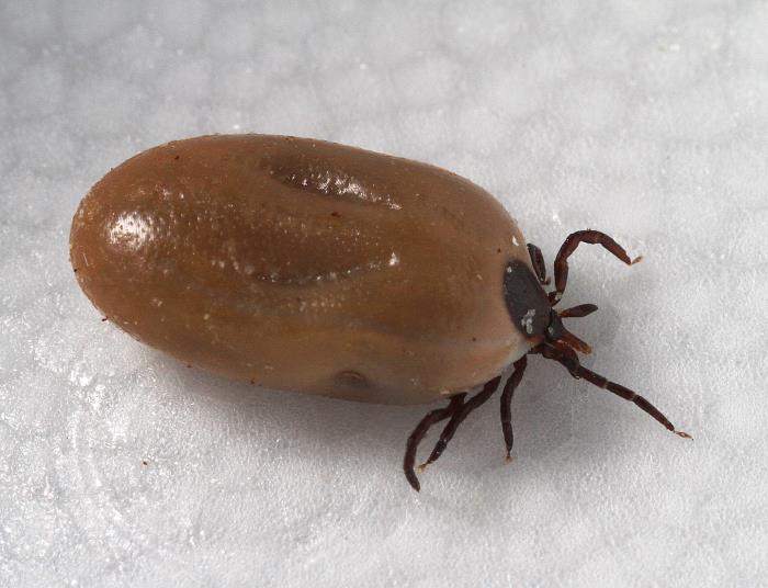

It is most common in the eastern and southern U.S. and as far north as Wisconsin and Maine.Īnaplasmosis: A rickettsial organism (Anaplasma phagocytophilum) spread through the bite of the black-legged (deer) tick and the brown dog tick. but is more common in Southern Africa.Ĭytauxzoonosis: A protozoan (Cytauxzoonosis felis) transmitted from Lone Star ticks, it’s often found in the southern U.S.

This disease is zoonotic, meaning it can be transmitted to people and should be reported to public officials.īabesiosis (Piroplasmosis): This is a protozoan (Babesia felis) that is spread through tick bites. Tularemia: A bacterium (Francisella tularensis) transmitted from the American dog tick and Lone Star tick often found everywhere in the U.S., except the Rocky Mountains and the Southwest. Hepatozoonosis: A protozoan that is spread through tick bites, this disease is not common in cats. Lyme disease: A bacterium (Borrelia burgdorferi) spread through the bite of the black-legged (deer) tick often found in the eastern U.S., and as far west as Texas and South Dakota. The six most common types of tick-borne diseases in cats include:

Most Common Types of Tick-Borne Disease in Cats
#Jackson lab tick borne disease doxy skin#
Ticks can be found anywhere in the United States and their bite can cause skin infections, anemia, and tick paralysis, which is a serious and life-threatening condition to cats. But did you know that Lyme disease can affect cats as well? Lyme Disease and many other diseases are spread through tick bites and are known as tick-borne diseases. Most people have heard of Lyme Disease, and unfortunately some may have suffered from the disease themselves. This mouse model of Lyme borreliosis, including the ability to monitor lesion development and spirochete load, can facilitate the testing of therapeutic regimens for the later stages of tick-transmitted Lyme disease and help investigate aspects of the immunopathogenesis of lesion development.The following content may contain Chewy links. N40-infected mice demonstrated a significantly higher spirochete load (an average of 1.23 spirochetes/mg of tissue, p = 0.045) in femorotibial joints 18 weeks after infection, with 60% of these mice maintaining lesions compared with those infected with B31 (13%), JD-1 (25%), or 910255 (50%), which averaged <0.5 spirochetes/mg of tissue. The amount of spirochetal DNA that could be amplified for heart at this time point was not statistically different between isolates, indicating a difference in virulence between B31 and N40 relative to JD-25. A higher percentage of B31- and N40-infected mice (92 and 100%, respectively) developed myocarditis than JD-1- or 910255-infected mice (67 and 46%, respectively) 12 weeks after tick bite. The q-PCR assay developed for this study was sensitive and could reliably measure as few as 1 to 10 spirochetes in the target tissues tested. Utilizing the Taqman quantitative polymerase chain reaction (q-PCR) fluorogenic detection technology to amplify a conserved region of the flagellin gene, a trend was demonstrated between the number of spirochetes in tissue with duration of pathology. In terms of the number of mice that demonstrated pathology in bladder, heart, and joint, the highest incidence of lesions occurred 12 weeks after tick bite. All isolates induced multifocal, lymphoid nodular cystitis, subacute, multifocal, necrotizing myocarditis, and a localized periostitis and arthritis of the femorotibial joint 6-18 weeks after tick infestation.

Four laboratory-grown, low-passage isolates of Borrelia burgdorferi sensu stricto, B31, JD-1, 910255, and N40, were incorporated into Ixodes scapularis ticks to examine the pathogenesis of these isolates in mice after tick transmission.


 0 kommentar(er)
